Advanced histological techniques for the study of Alzheimer’s disease pathophysiology.
Date and Time
Location
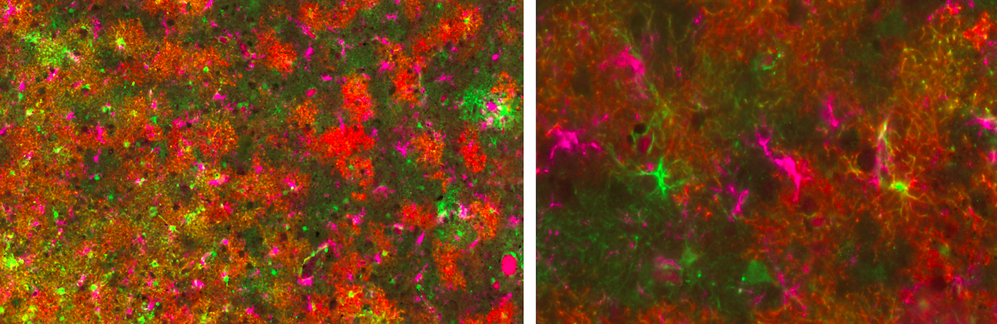
Alzheimer’s disease is the most common dementia in the elderly, affecting more than 5 million Americans. Pathologically, the Alzheimer’s disease brain is characterized by the deposition of the amyloid beta peptide in extracellular amyloid plaques and of the microtubule-associated protein tau in intraneuronal neurofibrillary tangles, together with severe loss of neurons resulting in brain atrophy. But, besides plaques, tangles, and atrophy, there is a prominent reaction of glial cells - astrocytes and microglia - which is gaining attention from the research community. The role of these glial responses in the pathophysiology of the disease remains uncertain, but some recent studies support a detrimental effect through either a gain of toxic function or a loss of neurotrophic support.