

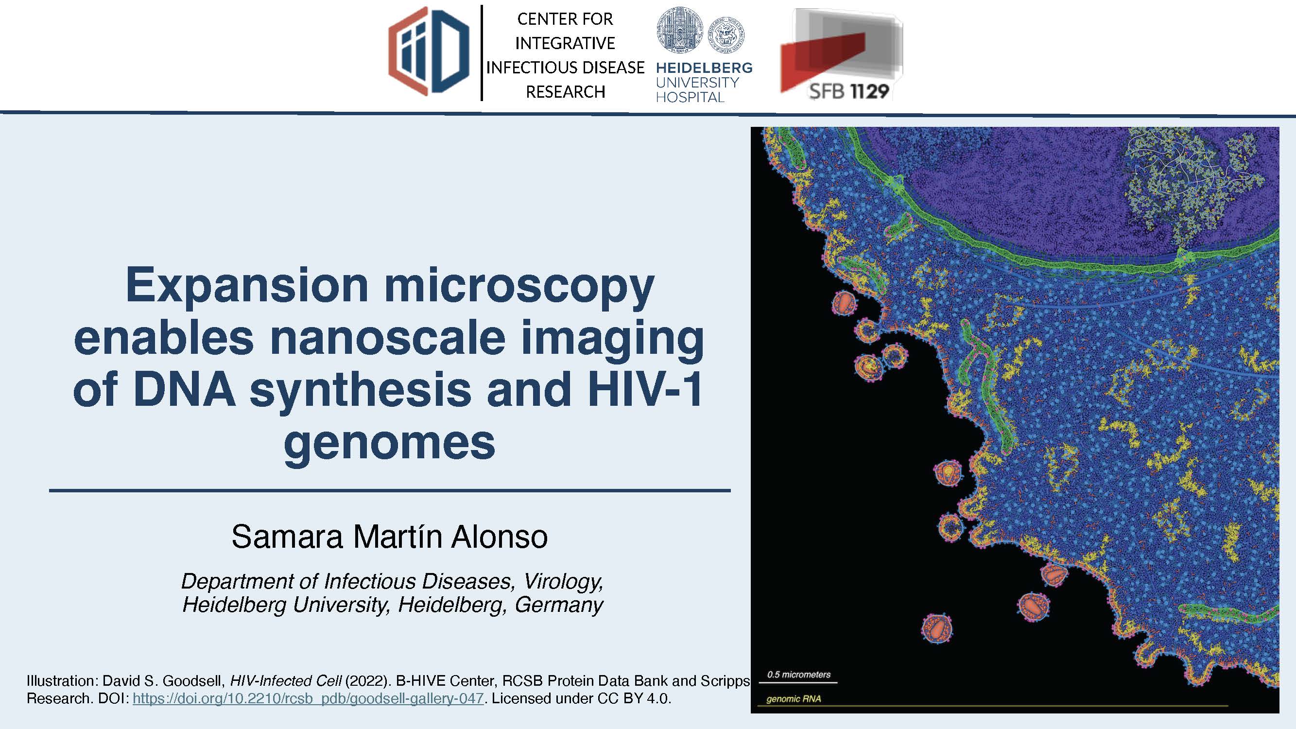
Expansion microscopy enables nanoscale imaging of DNA synthesis and HIV-1 genomes

Date and Time
The diffraction limit of conventional fluorescence microscopy restricts the visualisation of nanoscale features involved in DNA synthesis and virus–host interactions. Here, we developed an expansion microscopy-based workflow combining 5-ethynyl-2′-deoxyuridine (EdU) incorporation with biotin–streptavidin-mediated signal amplification for nanoscale detection of newly synthesised DNA following ultrastructure expansion microscopy (U-ExM). The method uses copper(I)-catalysed click chemistry to conjugate biotin to incorporated EdU, followed by streptavidin-based detection after expansion. We validated the approach by examining nuclear DNA replication throughout S-phase and mitochondrial DNA synthesis within individual nucleoids. We then applied it to primary human monocyte-derived macrophages (MDMs) infected with a reverse transcription-competent, non-infectious HIV-1 variant. In infected MDMs, U-ExM revealed discrete nascent HIV-1 DNA signals within nuclear speckles, spatially associated with clusters of cleavage and polyadenylation specificity factor 6 (CPSF6), a host factor that interacts with the HIV-1 capsid and contributes to viral nuclear trafficking. Nanoscale resolution further enabled the distinction of individual CPSF6-positive structures within these clusters. Following expansion correction, their dimensions were consistent with the reported size range of HIV-1 capsids, suggesting they may represent viral core-associated complexes. Overall, this workflow provides a versatile approach to investigate newly synthesised DNA and its spatial relationship with cellular structures and host factors, offering new opportunities to explore HIV-1 DNA organisation within the nucleus.
Speaker: Samara Martin Alonso (Department of Infectious Diseases, Virology,
Heidelberg University, Heidelberg, Germany)